Project Lay Summary:
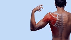
The word ‘dyskinesis’ is adapted from two words: ‘dys’ meaning bad/difficult and ‘kinesis’ meaning motion. Hence, Scapular Dyskinesia (SD) is an alteration in the normal resting position or active moving position of the shoulder blade (scapula) during shoulder movements. It is often associated with various shoulder conditions and commonly caused by a disturbance in muscle performance. Muscle performance is regulated by a muscle’s activation, strength and fatigue (repeated and intense use that causes a decline in muscle’s ability to generate force). House painters due to their repeated overhead arm posture and work requirements undergo easier fatigue of shoulder muscles which over a prolonged period of time affects their strength and joint motion. Pre-existing literature focuses on the alteration in scapular movement patterns caused due to fatigue and muscle weakness but activation remains less explored. Therefore, this study aims to analyse the scapular muscle activity in this at-risk population of house painters.
The participants will be approached through painting and decoration companies and associations. The study will be amongst 50 participants who will be divided into 2 groups based on the of presence or absence of SD. The study aims to calculate the muscle activity of the main scapular muscles, namely serratus anterior, upper, middle and lower trapezius and infraspinatus of both right and left sides using electric currents through an instrument called an electromyograph. After dividing the participants into groups, a base value muscle activity will be recorded while the muscles perform their maximum contraction against a device called a handheld dynamometer. This base value will be merged with the value recorded while the muscles perform a series of movements designed to fatigue them. This will form a total muscle activity of a particular muscle which will then be compared between the two groups. This will help to find out which muscle’s improper activity is related to SD in one group and if is it functioning normally in another group which will be beneficial to improve the SD treatment protocol in the future.
Background of the study:
The second international consensus conference on the scapula held in Lexington Kentucky concluded that though the exact role of SD in causing shoulder dysfunction remains unclear it has a high presence in most shoulder pathologies, particularly shoulder impingement. (Kibler et al.,2013) SD is often considered as an impairment to shoulder function and its proper evaluation can make the treatment strategies more effective. (Kibler et al., 2013) To improve treatment protocols it is important to know about the causes of SD. The causes of SD can be bone or joint-related, soft tissue related or neurological. (Giuseppe et al., 2020) Studies further categorised the factors causing SD as proximal which included muscle imbalances and distal which included internal imbalance like labral tears, GH instability, and acromioclavicular separation. (Kibler et al.,2013) Muscle imbalances imply defect in muscle activaction pattern, fatigue, strength or flexibility. The improper pattern of muscle activation and force of contraction as well as increased fatigue disturbs scapulohumeral rhythm leading to dyskinesia (Panagiotopoulos and Crowther, 2019). Current concepts of SD revolve only around the strengthening of scapular muscle. There is a need to shift the focus training of overall motor control of the scapula. (Sciascia and Kibler 2022) Furthermore, Panagiotopolus and Crowther (2019) also quote “the scapular musculature requires re-orientation in order to re-engage the correct pattern of activation.”.
House painters are a group of construction workers with a high prevalence of occupational musculoskeletal disorders specifically shoulder, neck and low back pain. (Pandey and Kiran, 2020) While supraspinatus tendinitis is a common condition seen in painters, rotator cuff tears are associated with long-term occupational load on their shoulders making them a vulnerable group for a probable shoulder injury. (Stenlund et al., 2002, Loew et al., 2019) A large variety of evidence shows repeated elevated arm activities increase shoulder muscle activity and shoulder load which in turn leads to easier muscle fatigue eventually causing shoulder pain and discomfort (Rosati et al. 2014). To critically appraise and find gaps in the available evidence of the current picture of SD and its treatment protocol in the painter population detailed search of the literature was done.
An in-depth search strategy was conducted on Pubmed, Scopus, EBSCO-Cinahl, OVID-Medline and Cochrane library databases. Articles were searched using the keywords mentioned in the table: 1. The key concepts of the search strategy were based on the population of painters, the intervention to be assessed being the muscle activity and the comparison based on SD. Synonyms and conceptual variants of these words were searched for using the Boolean operator ‘OR’ and these concepts were combined using the Boolean operator ‘AND’. The search was restricted to the English language and from the year 1996-2022 to maintain relevance. The discovered articles were managed through the Endnote 20 software where duplicates were removed. The remaining articles were shortlisted based on title and abstract relevant to the topic. Shortlisted articles were read through full-text links to find the close relevance to the key concepts mentioned earlier and the methodology used. (Flowchart 1) Lastly, the articles were critically appraised using the appropriate Critical appraisal skills program (CASP) checklist.
Table 1: Search terms, databases and results
Search terms | Search database | Total results | Number of articles after removing duplicates |
("Muscle activity" OR fatigue OR strength OR electromyography) AND ("Muscle activity" OR fatigue OR strength OR electromyography) AND ("SD" OR “scapular dyskinesis test” OR scapula OR shoulder)
| PUBMED | 123 | 123 |
SCOPUS | 204 | 118 | |
EBSCO-CINAHL | 126 | 26 | |
OVID- MEDLINE and AMED | 434 | 59 | |
COCHRANE LIBRARY | 36 | 15 |
Flowchart 1: Search strategy

Lopes et al. (2015) conducted a cross-sectional study to analyse the differences between scapular kinematics and shoulder muscle activity in patients of subacromial impingement syndrome with (n=19) or without (n=19) visual scapular SD. Two observers standardized to assessment and blinded by each other’s results but not by the shoulder pathology of the participants were appointed to divide the participants into a group of obvious SD and normal scapular motions using the scapular dyskinesis test (SDT). Scapular kinematics was analysed using an electromagnetic motion capture system. Upper, middle and lower trapezius, serratus anterior and infraspinatus muscle activity was studied using surface electromyography during ascending and descending phases of weighted shoulder flexion. Resisted muscle contraction prolonged for five seconds at the midpoint of the testing position was considered as a reference instead of maximum voluntary contraction for normalization due to the pain. The study concluded that the patients with obvious SD had less scapular external rotation and higher upper trapezius activity. The study could not confer whether the increased upper trapezius activity was in an attempt to control dyskinesia or was a cause of SD. The study highlighted that training of motor control and improving performance techniques to correct muscle imbalances may serve to reduce dyskinesia. Muscle activation and deactivation time as well as muscle latency should be considered for future research according to the authors
Plummer et al. (2017) in their study to categorize the prevalence of SD in participants with and without shoulder pain and relation to blinding of the pain factor in the examiner concluded that the prevalence reported by the unblinded examiner was higher when testing the involved shoulder for SD by SDT. This might have caused an experimenter bias reducing the internal validity of the study by Lopes et al. (2015) Another factor that might have led to a bias is that the participants only performed five bilateral weighted flexions whereas the SDT involves the performance of five bilateral weighted abduction along with flexion. This might have reduced the amount of fatigue in the shoulder muscles of participants with normal scapular motion which instead could have shown subtle or obvious SD. (McClure et al. 2009) The use of muscle contraction which was not maximal voluntary contraction to normalize the EMG might be accounted for a bias as it restricts the ability to compare between a group of individuals, different muscles in the same individual or different muscle groups. The use of submaximal muscle contraction could have been a more relevant choice (Halaki and Ginn, 2012)
Joshi et al. (2011) conducted a study to examine the effect of fatigue of shoulder external rotators on muscle activation and scapular kinematics in overhead athletes. In total, 25 participants without any history of shoulder pain participated in the study. The fatigue protocol used was weighted movements of the upper limb in proprioceptive neuromuscular facilitation (PNF) D2 pattern followed by fatigue for external rotators in the prone position. Surface electromyography was used to assess the muscle activity of the upper, middle and lower trapezius, serratus anterior and infraspinatus. Results were calculated using a Pearson product-moment correlation coefficient. It concluded that there was an increase in infraspinatus activity along with a decrease in lower trapezius activity. (r=–0.43, P=.04) Whereas, serratus anterior and upper trapezius activity remained unaffected by the fatigue protocol as these muscles were not fatigued sufficiently to affect their muscle activation. Authors suggested that the increased infraspinatus activity may be a compensatory mechanism to maintain the production of force despite of altered scapular position.
Ebaugh et al. (2006) conducted a similar study on 20 subjects without any history of shoulder pathology to determine the effect of shoulder external rotation muscle fatigue on the glenohumeral and scapulothoracic kinematics. Results showed a reduced external rotation of the humerus and a decrease in the posterior tilt of the scapula in the initial phase followed by more upward rotation in the middle ranges of arm elevation. When compared to Joshi et al. (2011) this study demonstrated a rise in the lower trapezius activity and a fall in the infraspinatus activity after an external rotation task. This difference may be due to the differences in the fatigue protocol as in the previous study the external rotators were fatigued at 90 degrees of glenohumeral abduction compared to 10 and 20 degrees in this study. Another difference was seen in the upper extremity tested. While the previous study tested only the dominant side this study tested both the dominant and nondominant sides. Yoshizaki et al. (2017) in their study identified differences in shoulder muscle activation between the dominant and non-dominant sides. One similarity between these two studies was that they collectively showed more upward rotation of the scapula after fatigue. This was challenged by a third study by Tsai et al. (2003) which reported a fall in the upward rotation of the scapula post-fatigue.
There is a conflict in the literature about the muscle activity of scapular muscles in the population with or without the presence of SD. Variability is seen in the fatigue protocol and the assessment measures using surface electrography. Fewer number of research and a variety of populations studied makes it more difficult to set a bias-free platform for comparison. Studies are conducted on the effect of fatigue and muscle weakness on scapular kinematics but the knowledge of specific activity patterns of scapular muscle in the painter population with visual SD remains unexplored. As per, the author’s knowledge there has been no similar study conducted on house painters. Further research is needed to evaluate the scapular muscle activity in this which may help to build on better prophylactic and therapeutic treatment for their shoulder-related issues.
Purpose of Study:
The study aims to analyse the difference in scapular muscle activity patterns in house painters with or without SD.
Objectives:
> To analyse if the post-fatigue muscle activity of serratus anterior, upper, middle, lower trapezius is greater or lesser in house painters with visual SD when compared to those who do not have SD.
> To analyse if the muscle activity of serratus anterior, upper, middle, and lower trapezius and infraspinatus is greater/lesser on the side of the shoulder with SD compared to the opposite side.
> To analyse if the muscle activity of the upper trapezius is greater in painters with SD compared to those without SD
> The muscle activity of the serratus anterior, middle and lower trapezius is lower in painters with SD compared to those without SD.
Null Hypothesis:
There is no difference in scapular muscle activity patterns in house painters with or without SD.
Alternate Hypothesis:
There is a difference in scapular muscle activity patterns in house painters with or without SD.
Research design and methods:
To analyse the differences in the muscle activity of scapular muscles in house painters with and without SD an observational case-control study is proposed. A case-control study is one where the researcher observes the factors linked to a disease or an outcome, where one group is the case group that has the disease or outcome and the other is a control group that is similar to the case group but does not have the disease or outcome of interest. (Tenny 2022) This study is proposed to use the case-control design as it studies the patterns of muscle activity in the same population with one significant difference being the presence of SD and not the characteristic of the population itself in which case it would have been a cross-sectional study.
If the outcome proposed (improper muscle activity) is found to be more commonly seen in the case group it can be hypothesized that there is an association between the outcome and the condition of the case group. (Tenny 2022) Although this study design has several advantages like it is less time-consuming when compared to a randomized control trial, helps to analyse rare diseases and conditions and helps to study multiple factors at once it also has certain disadvantages. It is difficult to get the control group exactly as similar to the case group giving a gateway to certain confounding biases. It can only be used to find an association and not causation of a condition. Another common disadvantage of this study is the potential recall bias but this study being conducted on an assessment basis instead of cognitive component rules out that factor.
Inclusion Criteria:
· Full-time working house painters with a working experience of at least 2 years (Stenlund et al., 2002)
· Age 20 to 50 years (as the prevalence of shoulder osteoarthritis increase after 50 years of age) (Juel and Natvig, 2014)
· Male (due to fewer number of women involved in the profession) (Stenlund et al., 2002)
· Working in Cardiff City
Exclusion Criteria:
· Shoulder pain
· History of any shoulder pathology in the last 2 years
· Restricted passive shoulder range of motion
· An overhead athlete (Harrison and Curtis, 2019)
· Active or passive cervical spine range producing shoulder symptoms (Harrison and Curtis, 2019)
· History of systemic musculoskeletal disease (Harrison and Curtis, 2019)
· History of a stroke causing functional impairment in the shoulder.
Sample Size:
A total sample of 50 participants is needed for this study with 25 in each group: case and control. This was proposed based on a priori power analysis on G*Power 3.1.9.7 and the parameters based on previous research. (Lopes et al., 2015, Kang, 2021)
The following parameters were used:
· Statistical t-test (mean: difference between two independent means)
· Two-tailed hypotheses was used based on the null hypothesis
· The effect size was 0.82 calculated from the research article of Lopes et al. (2015) using the Sensitivity calculation feature of G*power.
· α error was considered 0.05
· Power (1-ꞵ) was taken as 0.8 which is necessary for clinical research (Kang,2021)
While collecting samples during data collection there is a chance of occurrence of disparity in the number of participants with and without SD or in their similarity of demographic data. In such case, a matched-pair analysis will be used to yield the maximum number of matched pairs from the available samples. Data from the remaining unmatched participants will not be used for analysis. (Hannah et al. 2017) The study design requires a one-time assessment and participation by the participants hence, the risk of dropout of participants is minimal.
Data Collection and Analysis:
A convenience sampling technique will be used to reach out to the target population. Painting and decorating companies in Cardiff city will be contacted. (Loew et al., 2019) Posters will be put in the locations conducting the meetings of the workers association. The Painters and decorating Association of UK (PDA) will be contacted. Painters will be approached with the plan of the study and whoever meets the inclusion criteria and agrees to give their consent will be invited for the testing procedure.
Demographic data of all participants including their name, age, hand dominance, working experience and working hours will be recorded. This will help match pairs and maintain homogeneity in both groups. To divide them into two groups participants will be required to perform SDT (SDT) which has shown moderate reliability and validity. (Tate et al., 2009, Christiansen et al., 2017) Participants will have to perform 5 repetitions of bilateral, weighted, active shoulder flexion and bilateral, weighted, active shoulder abduction each to a 3-second count. The weight will be in the form of dumbbells of 1.4 kilograms (3lb) each for body weight less than 68.1 kilograms (150 lbs) and 2.3 kilograms (5lb) each for those weighing 68.1 kilograms or more. (McClure et al. 2009) Participants will be categorized as normal, subtle or obvious depending on the presence of dysrhythmia or winging of the scapula. (McClure et al. 2009) Participants with subtle dyskinesis will be excluded to maximize the potential for detecting between-group differences. (Lopes et al., 2015)
Muscle activity will be assessed by surface electromyography (EMG). The electromyograph will be connected to a laptop computer, where the EMG signals will be analysed using computer software like MATLAB/Naroxan depending upon the unit used for testing. EMG activity of the right and left serratus anterior, upper, middle and lower trapezius will be assessed as they are responsible for scapular motions. The activity of infraspinatus will also be assessed because of its prominent activity in repeated elevation and external rotation tasks. (Joshi et al., 2011) The placement of electrodes for these muscles is described in table 2. The area of the skin will be abraded if necessary and cleaned with isopropyl alcohol before placing the electrodes. Maximal voluntary isometric contractions (MIVC) will be performed for each muscle using the handheld dynamometer. Positions for these are described in table 3. The participant will be instructed to apply their maximum force for 5 seconds while measuring the MIVC for a specific muscle. Three trials of each muscle will be taken with 30 seconds rest between each trial and 1-minute rest between different muscles. (Joshi et al. 2011) The root mean square of MVIC will be used for the normalization of post-fatigue values. (Halaki and Ginn, 2012) Participants will then perform a fatigue protocol of 30 repetitions of weighted shoulder flexion with the same dumbbells used to test SDT. (Andres et al., 2019) A mark of standard complete overhead elevation will be maintained by asking the participants to touch their ears with their arms during elevation. Speed will be maintained by a metronome of 20 beats per minute. The root mean square of the EMG activity while performing the protocol will be calculated for the specific muscles and normalized against the MIVC to be used for data analysis.
Table 3: Placement of EMG electrodes (Lopes et al., 2015)
Muscle | Electrode Placement |
Serratus anterior | Along the midaxillary line over the 6th rib |
Upper trapezius | Immediately lateral to a point midway between the spinous process of T1 and the acromion process |
Middle trapezius | Immediately lateral to a point midway between the spinous process of T3 and the root of spine of the scapula |
Lower trapezius | Immediately lateral to a point midway between the spinous process of T7 and the inferior angle of the scapula |
Infraspinatus | 2.54cm inferior to the spine of the scapula at a point midway between the root of the spine of the scapula and the posterior acromion process |
Table 4: Position to test the MIVC (Joshi et al., 2011)
Muscles | Position for testing MIVC | HHD placed | Manual Resistance applied |
Serratus anterior | Sitting with the arm abducted to 125degrees | Above the elbow | To the inferior angle of the scapula, in an attempt to de-rotate it. |
Upper trapezius | Sitting with the arm abducted to 90 degrees with the neck bent to the same side, rotated to the opposite side and extended | Above elbow with the force in the direction of adduction | To the head directed towards the neutral position |
Middle trapezius | Prone with the arm abducted to 90 degrees | Distal to the elbow joint | Distal to the elbow joint in the direction perpendicular to the floor |
Lower trapezius | Prone with the arm abducted to 125 degrees | Distal to the elbow joint | Distal to the elbow joint in the direction perpendicular to the floor |
Infraspinatus | Prone with arm externally rotated in 90 degrees abduction | Proximal to the wrist joint | To the distal end of the forearm in the direction of internal rotation |
Data analysis will be done using the ‘SPSS software for Windows’. To adjust the comparison between multiple muscle groups Bonferroni correction will be used. (Haynes, 2013) The data collected in this study will be the odd ratio. Chi-Square test will be used to find the difference level in the muscle activity of the particular muscles in both groups. The significance of differences between the two groups will be compared with the past literature to find its confidence level. The conclusion and implications of the study will then be based on the strength of the link between outcome and exposure.
Ethical Consideration:
In accordance with the Cardiff University Research Integrity and Governance Code of Practice (2019), ethical considerations for the conduction of this study will be made. Firstly, ethical approval will be obtained from the Cardiff University School of Healthcare Science research and ethics committee. Thereafter, an application will be made to the approaching house painting company’s owner/ manager to seek their ethical approval. Although, asking participants to devote their time to participate in the study is not ideal. They will be explained the aims of the study and the potential benefits for them and their colleagues keeping in mind the principle of beneficence (Gelling 1999). They will also be directed to appropriate referrals and services necessary for the rehabilitation if they consent to it.
Informed consent of human participants is essential according to the Nuremberg Code of 1947 (1996) Informed written consent will be taken from the participants after providing a sheet of detailed information about the research. The information sheet will contain the aim, objectives, information about the test procedure, what participants will be needed to do, how the data will be stored, how the data will be managed and used for analysis, how long will the data be stored and used for publication or further research. Appointments for the test procedure will be arranged according to the participant’s convenience and the availability of the laboratory setup. All participants will be treated equally abiding by the principle of justice (fair and equitable) (Gelling 1999) Participants can choose to withdraw their consent or decline the use of the data at any point during the research giving them the right of the principle of Autonomy. (Gelling 1999)
Patient safety will be considered at every point of the test process. Both physical and mental inhibition of the patient regarding the risk and harms of an electrodiagnostic test will be kept in mind. Participants will be explained the use of surface electromyography and its harmless effects following the principle of non-maleficence (Gelling 1999) Consent for performing the use of the surface electrodiagnostic test will be included in the written consent. Facilities for emergency action if the participant becomes unwell at any point during the testing procedure will be arranged.
Collection, storage and protection of data will be followed according to Data Protection Act 1998. All patient information will be stored digitally without any hard copies. The anonymity and confidentiality of the participants will be maintained while storing and using the data. (Bos, 2020) No participant’s identification data will be disclosed anywhere in the database. All participants will be allocated numbers against which their demographic data will be stored. This data along with the study data will be protected by passwords and kept on the researcher’s computer which will be protected by a password as well. The participant’s demographic information and study data will be then stored on the Cardiff University research records according to Cardiff University Records Management Policy (2015) Participants will be informed about the time duration for which the records will be held before discarding them.
Dissemination:
The study findings will be shared by the participants and clinical settings. A summary or poster presentation of the study will be shared with the companies recruiting house painters. Information about the changes in the work techniques, duration of work, work equipment, work setup, ergonomic consideration and prophylactic and therapeutic training of house painters will be given to the companies based on of previous literature review. The findings will be shared with the Painting and decorating Association PDA which is the United Kingdom’s (UK) largest membership association representing painting and decorating businesses in a hope of taking necessary changes to reduce the risk factors associated with this occupation and eventually prolonging their retirement age. It will also be shared in the physiotherapeutic clinical settings and hospitals to treat such patients efficiently. It will be up for publication and be presented at conferences where this issue can be discussed at a greater level to compare these findings elsewhere in the UK.
References:
Andres, J., Painter, P.J., McIlvain, G. and Timmons, M.K. (2019). The Effect of Repeated Shoulder Motion on Scapular Dyskinesis in Army ROTC Cadets. Military Medicine 185(5-6), pp. e811–e817. doi: 10.1093/milmed/usz408
Bos, J. (2020). Confidentiality. Research Ethics for Students in the Social Sciences, pp. 149–173. [Online article] Available at: https://link.springer.com/chapter/10.1007/978-3-030-48415-6_7 [Accessed: 8 January 2023].
Christiansen, D.H., Møller, A.D., Vestergaard, J.M., Mose, S. and Maribo, T. (2017). The scapular dyskinesis test: Reliability, agreement, and predictive value in patients with subacromial impingement syndrome. Journal of Hand Therapy 30(2), pp. 208–213. doi: 10.1016/j.jht.2017.04.002
Critical Appraisal Skills Programme (2018). CASP Checklist. Available at: https://casp-uk.net/casp-tools-checklists/ [Accessed: 6 January 2023]
Data Protection Act 1998. (2023). Available at: https://www.legislation.gov.uk/ukpga/1998/29/contents [Accessed: 4 January 2023].
Gelling, L. (1999). Ethical principles in healthcare research. Nursing Standard 13(36), pp. 39–42. doi: 10.7748/ns1999.05.13.36.39.c2607.
Giuseppe, L.U. et al. (2020). Scapular Dyskinesis: From Basic Science to Ultimate Treatment. International Journal of Environmental Research and Public Health, [online] 17(8). doi:10.3390/ijerph17082974.
Halaki, M. and Gi, K. (2012). Normalization of EMG Signals: To Normalize or Not to Normalize and What to Normalize to? Computational Intelligence in Electromyography Analysis - A Perspective on Current Applications and Future Challenges. doi: 10.5772/49957
Hannah, D.C., Scibek, J.S. and Carcia, C.R. (2017). Strength Profiles in Healthy Individuals with and Without Scapular Dyskinesis. International journal of sports physical therapy 12(3), pp. 305–313.
Harrison, N. and Curtis (2019). The Effect of Serratus Anterior Fatigue on Scapular Kinematics. [Online article] Available at: https://mds.marshall.edu/ctd [Accessed: 8 January 2023].
Haynes, W. (2013). Bonferroni Correction. Encyclopedia of Systems Biology , pp. 154–154. DOI: 10.1007/978-1-4419-9863-7_121
Joshi, M., Thigpen, C.A., Bunn, K., Karas, S.G. and Padua, D.A. (2011). Shoulder External Rotation Fatigue and Scapular Muscle Activation and Kinematics in Overhead Athletes. Journal of Athletic Training 46(4), pp. 349–357. doi: 10.4085/1062-6050-46.4.349
Juel, N.G. and Natvig, B. (2014). Shoulder diagnoses in secondary care, a one year cohort. BMC Musculoskeletal Disorders 15(1). doi:https://doi.org/10.1186/1471-2474-15-89
Kang, H. (2021). Sample size determination and power analysis using the G*Power software. Journal of Educational Evaluation for Health Professions 18, p. 17. doi: 10.3352/jeehp.2021.18.17
Kibler, W.B., Ludewig, P.M., McClure, P.W., Michener, L.A., Bak, K. and Sciascia, A.D. (2013). Clinical implications of scapular dyskinesis in shoulder injury: the 2013 consensus statement from the ‘scapular summit’. British Journal of Sports Medicine, [online] 47(14), pp.877–885. doi:10.1136/bjsports-2013-092425.
Loew, M., Doustdar, S., Drath, C., Weber, M.-A., Bruckner, T., Porschke, F., Raiss, P., Schiltenwolf, M., Almansour, H. and Akbar, M. (2019). Could long-term overhead load in painters be associated with rotator cuff lesions? A pilot study. PLOS ONE, 14(3), p.e0213824. doi: 10.1371/journal.pone.0213824.
Lopes, A.D., Timmons, M.K., Grover, M., Ciconelli, R.M. and Michener, L.A. (2015). Visual Scapular Dyskinesis: Kinematics and Muscle Activity Alterations in Patients With Subacromial Impingement Syndrome. Archives of Physical Medicine and Rehabilitation 96(2), pp. 298–306. doi: 10.1016/j.apmr.2014.09.029
McClure, P., Tate, A.R., Kareha, S., Irwin, D. and Zlupko, E. (2009). A Clinical Method for Identifying Scapular Dyskinesis, Part 1: Reliability. Journal of Athletic Training 44(2), pp. 160–164. doi: 10.4085/1062-6050-44.2.160
Panagiotopoulos, A.C. and Crowther, I.M. (2019). Scapular Dyskinesia, the forgotten culprit of shoulder pain and how to rehabilitate. SICOT-J, 5, p.29. doi:10.1051/sicotj/2019029.
Pandey, P. and V. Kiran, U. (2020). Postural Discomfort and Musculoskeletal Disorders among Painters - An Analytical Study. Journal of Ecophysiology and Occupational Health, 20(3&4), pp.203–208. doi:10.18311/jeoh/2020/25320.
Plummer, H.A., Sum, J.C., Pozzi, F., Varghese, R. and Michener, L.A. (2017). Observational Scapular Dyskinesis: Known-Groups Validity in Patients With and Without Shoulder Pain. Journal of Orthopaedic & Sports Physical Therapy 47(8), pp. 530–537. doi: 10.2519/jospt.2017.7268.
Rosati, P.M., Chopp, J.N. and Dickerson, C.R. (2014). Investigating shoulder muscle loading and exerted forces during wall painting tasks: Influence of gender, work height and paint tool design. Applied Ergonomics 45(4), pp. 1133–1139. doi:10.1016/j.apergo.2014.02.002
Sciascia, A. and Kibler, W.B. (2022). Current Views of Scapular Dyskinesis and its Possible Clinical Relevance. International Journal of Sports Physical Therapy. 17(2):117-13 doi:10.26603/001c.31727.
Stenlund, B., Lindbeck, L. and Karlsson, D. (2002). Significance of house painters’ work techniques on shoulder muscle strain during overhead work. Ergonomics, 45(6), pp.455–468. doi:10.1080/00140130210136954.
Tate, A.R., McClure, P., Kareha, S., Irwin, D. and Barbe, M.F. (2009). A Clinical Method for Identifying Scapular Dyskinesis, Part 2: Validity. Journal of Athletic Training 44(2), pp. 165–173. doi: 10.4085/1062-6050-44.2.165
Tenny, S., Kerndt, C.C. and Hoffman, M.R. (2022). Case Control Studies. [Online article] Available at: https://www.ncbi.nlm.nih.gov/books/NBK448143/ [Accessed: 6 January 2023].
The Nuremberg Code (1947). (1996). BMJ 313(7070) [Online] Available at: https://doi.org/10.1136/bmj.313.7070.1448 [Accessed: 6 January 2023]
Tsai, N.-T., McClure, P.W. and Karduna, A.R. (2003). Effects of muscle fatigue on 3-dimensional scapular kinematics11No commercial party having a direct financial interest in the results of the research supporting this article has or will confer a benefit upon the author(s) or upon any organization with which the author(s) is/are associated. Archives of Physical Medicine and Rehabilitation 84(7), pp. 1000–1005. doi: 10.1016/s0003-9993(03)00127-8.
Yoshizaki, K., Hamada, J., Tamai, K., Sahara, R., Fujiwara, T. and Fujimoto, T. (2009). Analysis of the scapulohumeral rhythm and electromyography of the shoulder muscles during elevation and lowering: Comparison of dominant and nondominant shoulders. Journal of Shoulder and Elbow Surgery 18(5), pp. 756–763. doi:10.1016/j.jse.2009.02.021


